Menu

FESEM | SEM5000 Pro

FESEM | SEM5000 Pro

High Resolution under Low Excitation

Product information

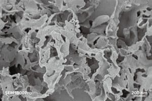
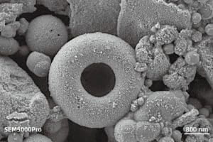
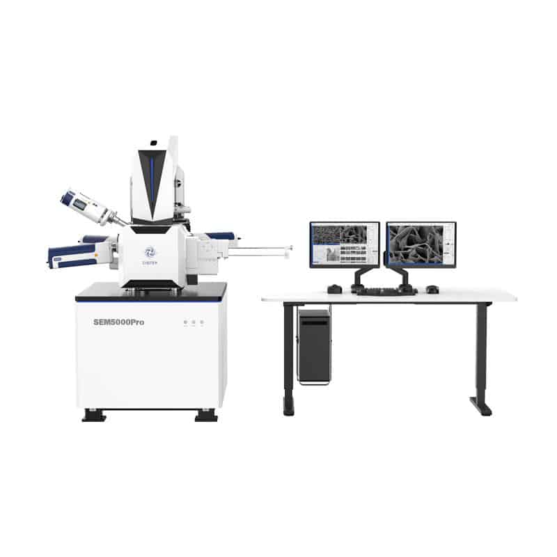
The CIQTEK SEM5000Pro is a Schottky field emission scanning electron microscope (FE-SEM) specialized at high resolution even under low excitation voltage. Employing an advanced “Super-Tunnel” electron optics technology facilitates a crossover-free beam path together with an electrostatic-electromagnetic compound lens design.

These advancements reduce spatial charging effect, minimize lens aberrations, enhance imaging resolution at low voltage, and achieve a resolution of 1.2 nm at 1 kV, which allows for direct observation of non-conductive or semi-conductive samples, effectively reducing sample irradiation damage.


Electron optics

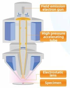
★ “Super Tunnel” electron optics column technology/in-lens beam deceleration
Decrease spatial charging effect, ensuring low voltage resolution.

★ Crossover-free in the electron beam path
Effectively reduce lens aberrations and improve resolution.

★ Electromagnetic & electrostatic compound objective lens
Reduce aberrations, significantly improve resolution at low voltages, and enable observation of magnetic samples.

★ Water-cooled constant-temperature objective lens
Ensure the stability, reliability, and repeatability of the objective lens operation.

★ Variable multi-hole aperture with electromagnetic beam deflection system
Automatic switching between apertures without mechanical motion, allowing fast switching between imaging modes.

Life Science

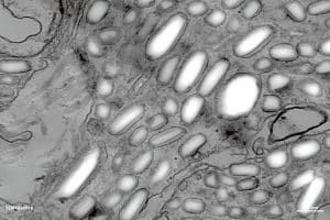
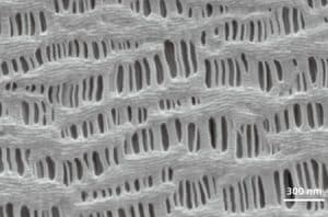
Characterization of iridophores in lizard skin cells, using the STEM detector in the CIQTEK SEM5000Pro FE-SEM.

Animal colors in nature can be classified into two categories based on their formation mechanisms: pigment colors and structural colors.
Pigment colors are achieved through variations in pigment composition and the overlapping of colors, similar to the principles of “primary colors.”
Structural colors, on the other hand, are generated through the reflection of light of different wavelengths by intricate physiological structures, based primarily on principles of optics. Iridophores, found in lizard skin cells, possess structures similar to diffraction gratings. We refer to these structures as “crystalline plates.” Crystalline plates can reflect and scatter light of different wavelengths. Studies have shown that by varying the size, spacing, and angle of the crystalline plates in lizard iridophores, the wavelengths of light scattered and reflected by their skin can be altered. This finding is significant for understanding the mechanisms behind color change in lizard skin.


Software options

  • Particle & Pore Analysis Software (Particle) *Optional
    The CIQTEK SEM Microscope software employs various target detection and segmentation algorithms, suitable for different types of particle and pore samples. It enables quantitative analysis of particle and pore statistics and can be applied in fields such as materials science, geology, and environmental science.
  • Image Post-processing Software *Optional
    Perform online or offline image post-processing on images captured by electron microscopes and integrate commonly used EM image processing functions, convenient measurement, and annotation tools.
  • Auto Measure *Optional
    Automatic recognition of line width edges, resulting in more accurate measurements and higher consistency. Support multiple edge detection modes, such as Line, Space, Pitch, etc. Compatible with multiple image formats and equipped with various commonly used image post-processing functions. The software is easy to use, efficient, and accurate.
  • Software Development Kit (SDK) *Optional
    Provides a set of interfaces for controlling the SEM microscope, including image acquisition, operating condition settings, power on/off, stage control, etc. Concise interface definitions allow for the rapid development of specific electron microscope operation scripts and software, enabling automated tracking of regions of interest, industrial automation data acquisition, image drift correction, and other functions. Can be used for software development in specialized areas such as diatom analysis, steel impurity inspection, cleanliness analysis, raw material control, etc.
  • AutoMap *Optional


Features

  • Low Voltage High Resolution

    In-lens electron detector image at 200 V low excitation voltage, achieves non-destructive morphological characterization of lithium-ion polymer membrane fiber structures.

  • BSED-based ECCI mode (Electron Channeling Contrast Imaging)The “Electron Channeling effect” refers to a significant reduction in electron scattering by crystal lattices, when the incident electron beam satisfies the Bragg diffraction condition, allowing a large number of electrons to pass through the lattice, thus exhibiting a “channeling” effect.For polycrystalline materials with uniform composition and polished flat surfaces, the intensity of backscattered electrons relies on the relative orientation between the incident electron beam and crystal planes. Grains with larger orientation variation exhibit stronger signals therefore brighter images, qualitative characterization with such grain orientation map is achieved.
  •  Simultaneously Multi-channel Imaging via Various Detectors
  • Retractable Scanning Transmission Electron Microscopy (STEM) Detector
  • High Stability
  • In-lens Electron Detector
  • *Specimen Exchange Loadlock (8 inches compatible)
  • Electromagnetic Beam Deflection with Multi-hole Apertursofte
  • Excellent Expandability

*optional


Image gallery

Non-conductive two-dimensional material C3N4, non-destructive characterization of its fine layer structure under low-voltage conditions at 500 V
Lithium-ion Battery Cathode Precursor – Iron Phosphate with poor conductivity, imaged using an In-lens Electron Detector (In-lens) under low voltage (1 kV) conditions
Polymer magnetic beads, with poor conductivity, characterized using a low-voltage at 2 kV with Everhart-Thornley Detector (ETD) for the characterization of biologically targeted drug delivery
Imaging of nickel foam with strong three-dimensional effect using a 2 kV excitation voltage with Everhart-Thornley Detector (ETD)
Polymer magnetic beads, characterized using a low-voltage 2 kV with Everhart-Thornley Detector (ETD), exhibit a strong sense of three-dimensionality

Downloads

No downloads found.